
Imaging
The secondary electron (SE) and backscattered electron (BSE) signals are typically used for sample imaging, with a much lesser use of characteristic X-rays (element mapping). The ESS JEOL 733 does not easily have the capability to image cathodoluminescence (CL).
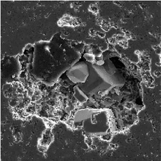
Secondary electron imaging (SE) is used primarily to image 3-dimensional objects or features. As the SE detector is positioned to the right side of the sample, surfaces or edges directed toward the detector will produce a higher signal (bright on the CRT screen) then those directed away from the detector (dark on the CRT screen). These ‘edge effects’ create the 3-D appearance.
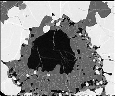
The number of electrons ‘backscattered’ from the sample (BSE) depends on the average atomic number (Z) of the target material. Thus, in multicomponent materials, like rocks, the individual minerals can be distinguished based on the fraction of electrons backscattered. Even variations in chemistry within a single mineral can be observed in BSE images.
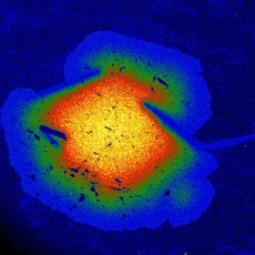
The 2-dimensional distribution of elements can be imaged by counting x-rays (instead of SE or BSE) as the primary beam is rastered across the sample. This example shows the distribution of Mn; the most intense the color the higher the concentration. The ESS JEOL 733 can collect a maximum of eight element x-rays at one time (4 WDS plus 4 EDS).
Mosaic Imaging
Although the minimum magnification of the JEOL 733 microprobe is 40x, it is possible to tile together 40x images using the GELLER software into a mosaic image of much lower magnification (ie., one can image larger areas). See an example, here.
click image
click image