Secondary Electrons

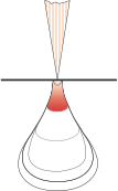
Secondary electrons are low energy electrons (<50eV) ejected from the conduction band of a sample following interaction with the high energy primary beam. Generally considered to be emitted from the upper <~100 nm of the analytical volume, they are thus well suited to image fine scale, three dimensional structures.

Comparison of BSE and SE imaging
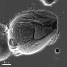
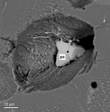
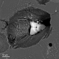
Note that the SEI image (below) has a 3-D look but there is no tonal differentiation between pyrrhotite (Fe, S) and clinopyroxene (Ca, Al, Ti, Mg, Fe, Si, O). A clear compositional distinction is seen in the BSE image, but the image is ‘flat’. These signals can be mixed on the microprobe CRT screen to display a 3-D compositional image.
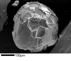
Smelter Glass Bead
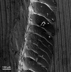
Groove In Brass
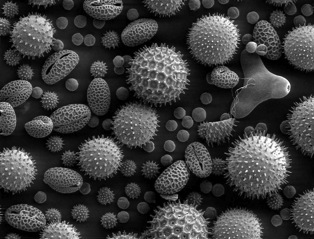
pollen (from web)
SEI image of pyrrhotite-bearing vug in clinopyroxene
BSE image of pyrrhotite-bearing vug in clinopyroxene
Mixed SEI + BSE image