BEE 531 Ultrasound Imaging
Instructor: Matthew Bruce / mbruce@uw.edu
University of Washington
Introduction to the course:
- My background
- Where does ultrasound fit in imaging?
- History of ultrasound
- Introduction to ultrasound system
- Course Overview
Background
What is Diagnostic imaging?
Detects injuries, diagnoses diseases, guides treatments and monitors conditions.
Where does ultrasound fit in diagnostic imaging?
- Other imaging modalities
- Advantages of US
- Disadvantages of US
- When is US used?
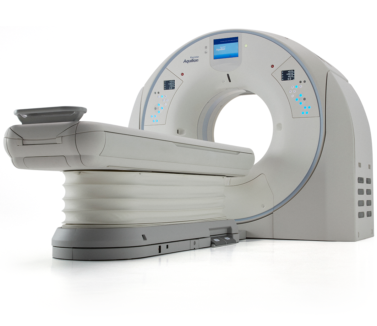
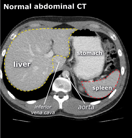
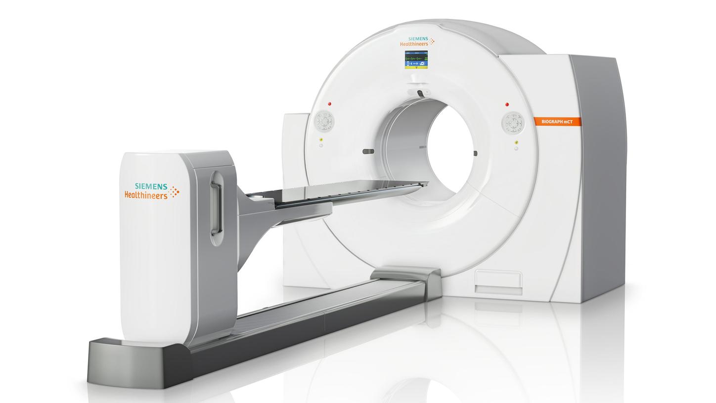
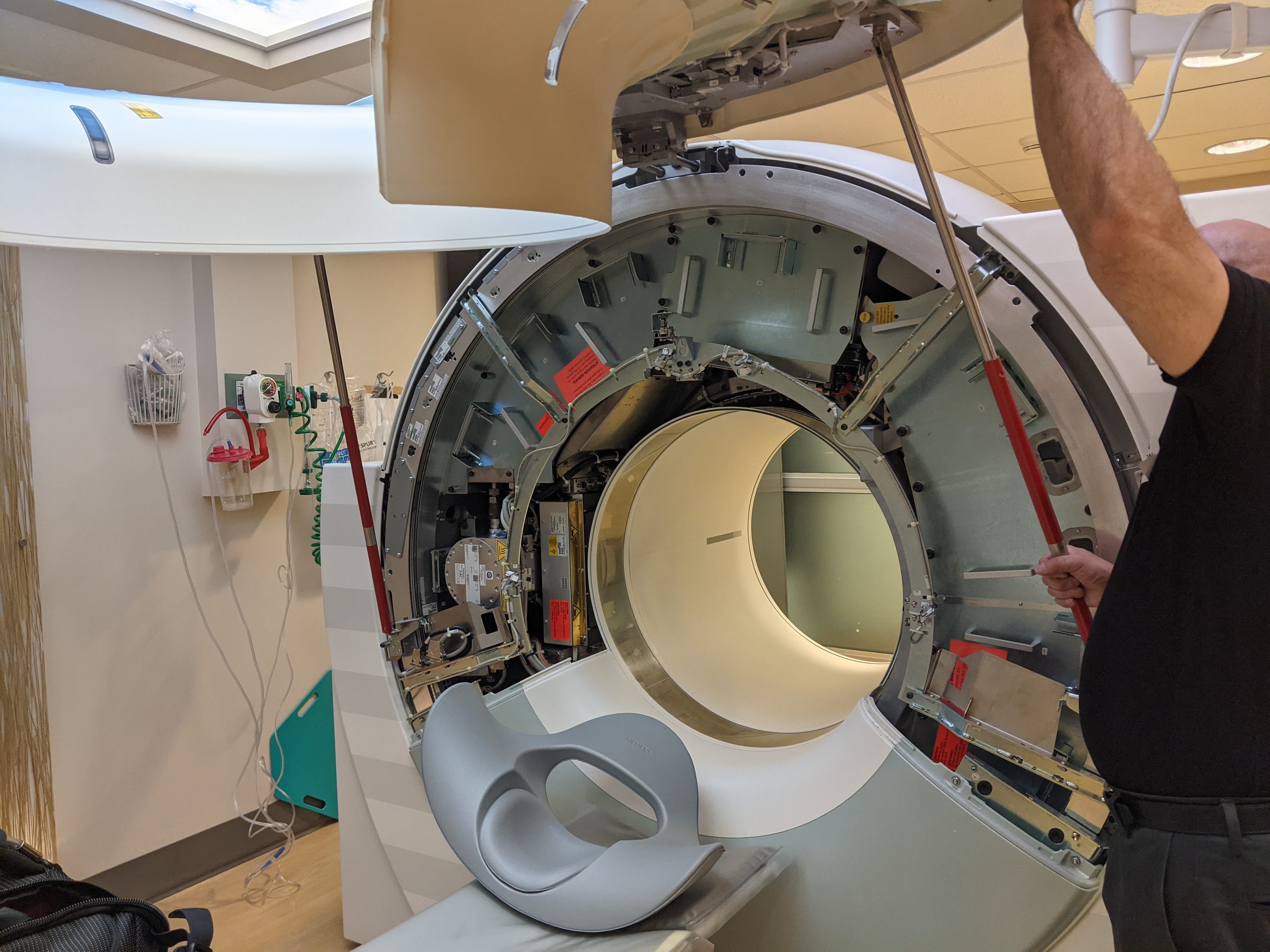
Computed x-ray tomography (CT)
Advantages
- Estimates x-ray absorption
- Access to volumes
- Widely available in certain areas.
Disadvantages
- Adds to cumulative lifetime radiation exposure
- Length of acquisition ~ 20 min (motion)
- Cost $400-700
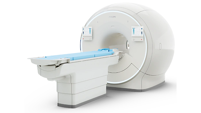
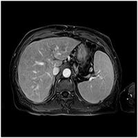
Magnetic Resonance Imaging
Advantages
- Estimates magnetic relaxation times (i.e. T1/T2).
- Access to volumes
- No radiation.
Disadvantages
- Availability ($1-3 million).
- Length of acquisition ~ 20 min (motion)
- Cost $700-1200
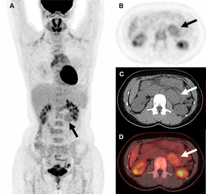
Nuclear - (includes PET)
Advantages
- Estimates cellular function/ activity.
- Access to volumes
- Very sensitive.
Disadvantages
- Radiation.
- Radiotracer short half lifes made in cyclotron (2 cyclotrons in South Seattle).
- Availability restricted.
- Length of acquisition/ resolution ~ 20 min (motion).
- Cost ~$300-3000 (PET $~6000
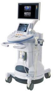
Ultrasound
Advantages
- Estimates backscatter of ultrasound waves.
- No radiation.
- Highly portable
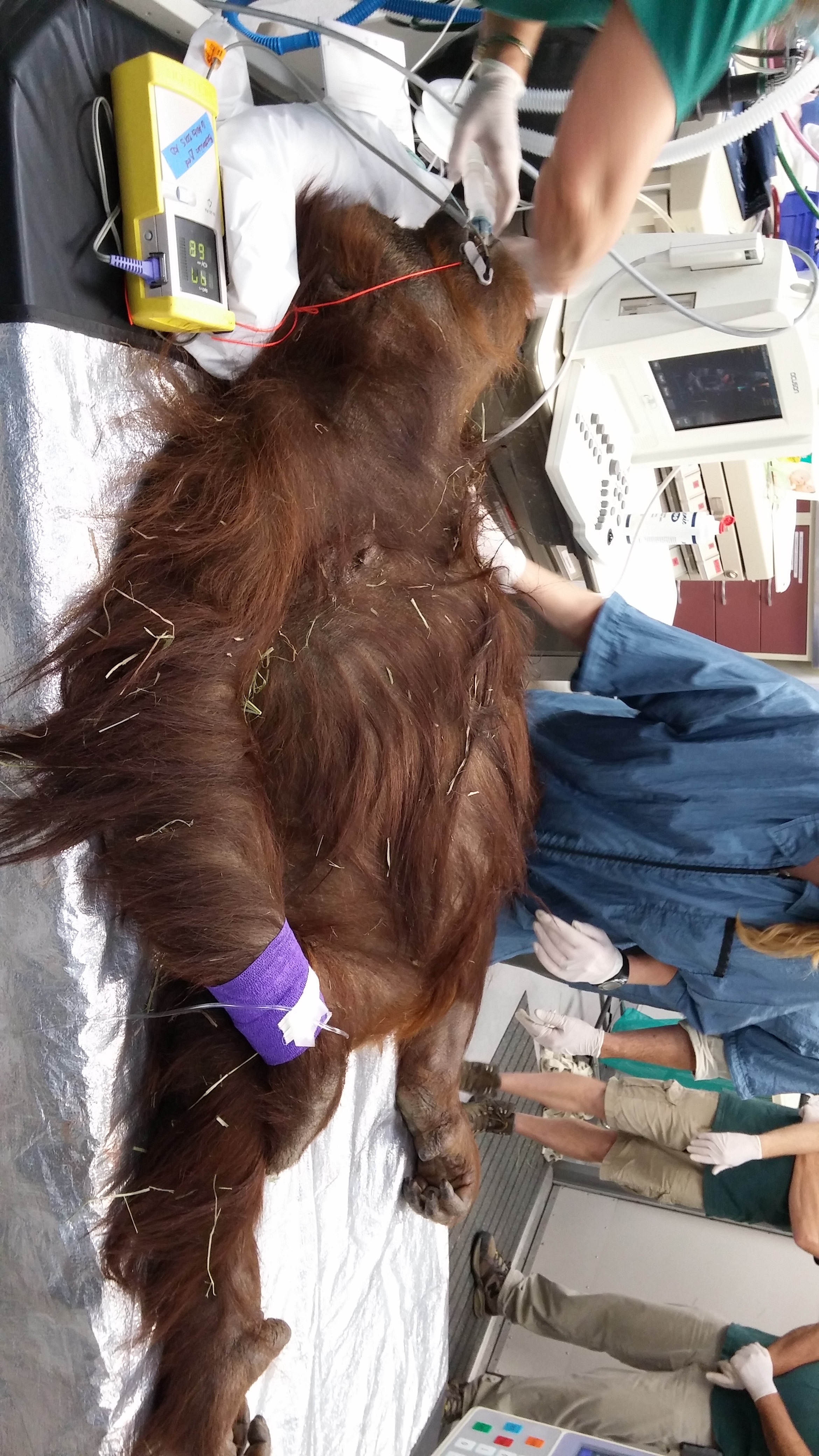
- Real time (used often to guide interventional procedures)
- Low cost $100-200
Disadvantages
- Limited access in some circumstances.
- Access to brain
- Difficult to access to abdominal organs.
- Limited access to volumes.
- Acoustically difficult patients.
- Cost $100-200
Why ultrasound?
- Low cost
- No radiation
- Non-invasive
- Highly portable
- Real-time imaging
Why I love ultrasound?
- Impacts lives of others
- Expanding applications user base/capabilities
- Lots of cool gadgets/technology (transducers, math, processing, computing/cpu/gpu
- Figure out new problems and how biological processes
Big shifts in Ultrasound imaging
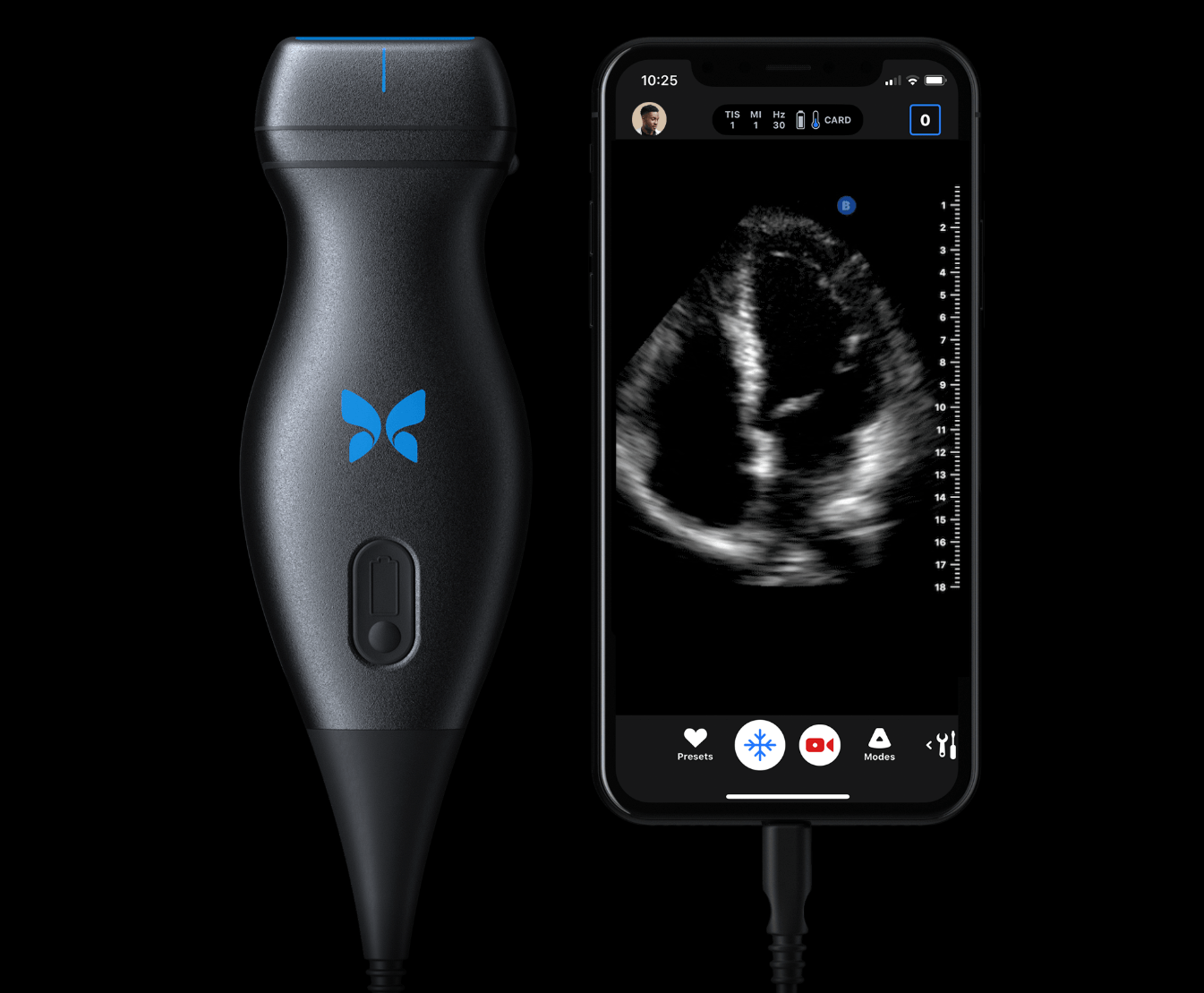
- Miniaturisation/portability pushing ultrasound into new places/uses
- Increase in computation (both gpu/cpu) enabling new opportunities
- New transducer technologies
- Artificial Intelligence
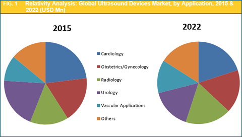
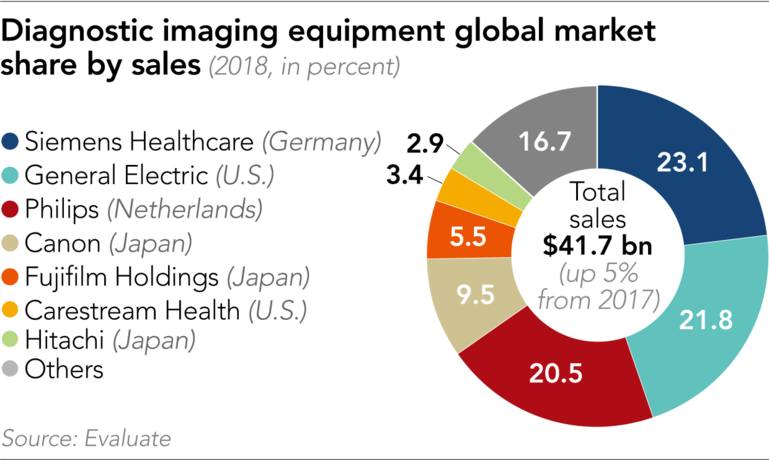
Ultrasound Market
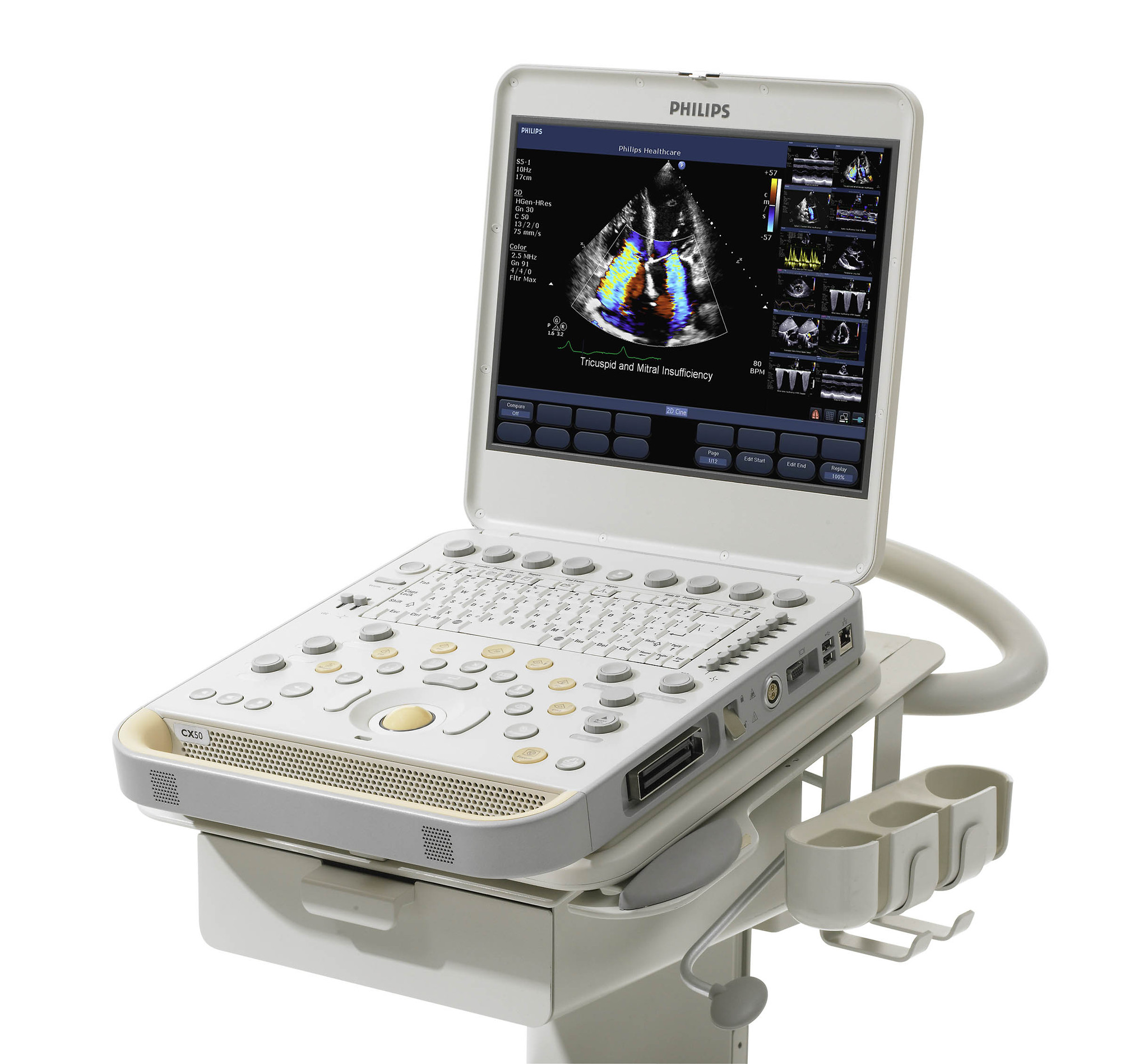
Pacific Northwest Ultrasound Companies
- Philips Medical Systems (Bothell)
- Siemens Ultrasound (Issaquah)
- Fujifilm/Sonosite (Bothell)
- Verasonics (Kirkland)
- EchoNous (Kirkland)
- Spencer Technologies (Redmond)
- United Imaging (Bellevue)
- Sonoscape (Kirkland)
- Sonic Concepts (Bothell)
- BioSound/Sonoscape (Redmond)
- Ekos/Boston Scientific (Bothell)
- Cerevast (Bothell)
- Mirabils Medica (Bothell)
- Verathon (Bothell)
- Clarius (Surrey)
- Wave (San Francisco)
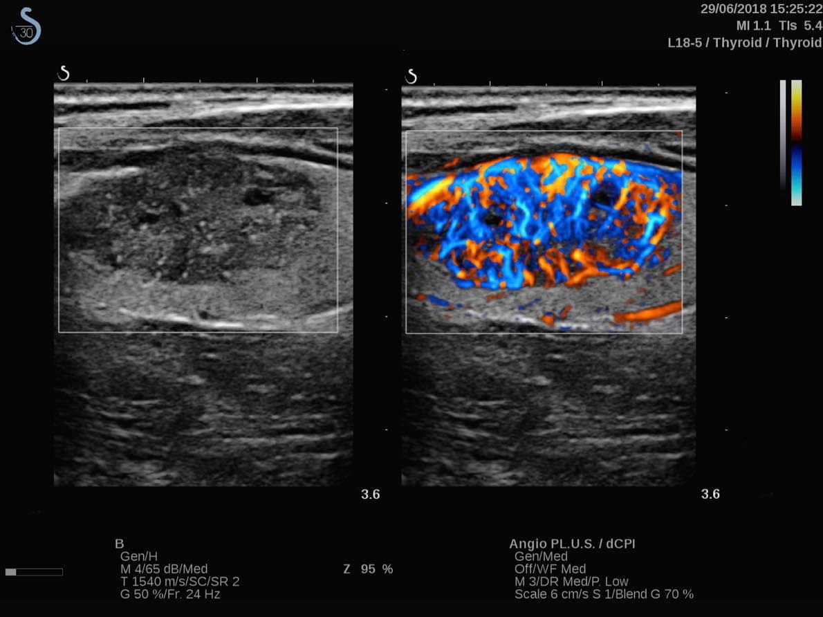
Cardiology-Echocardiology
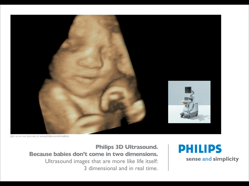
Obstetrics/Gynecology
Obstetrics/Gynecology
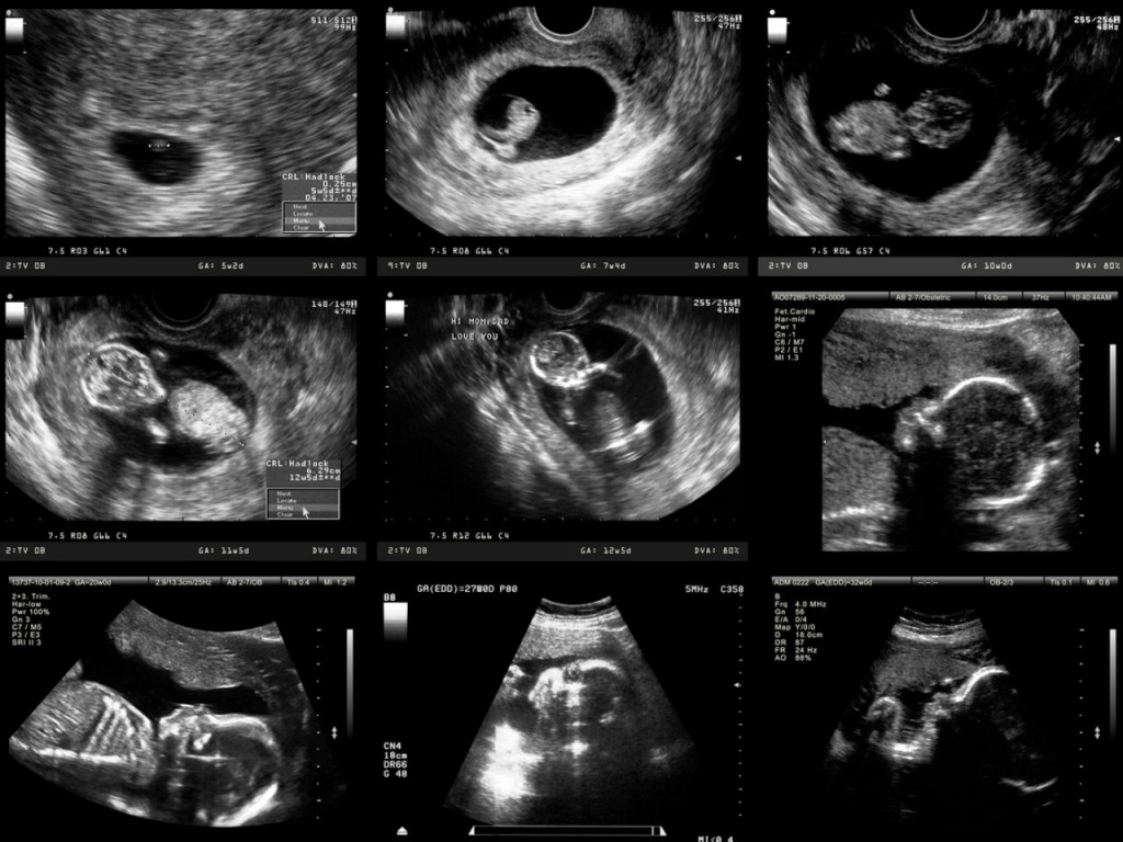
Obstetrics/Gynecology
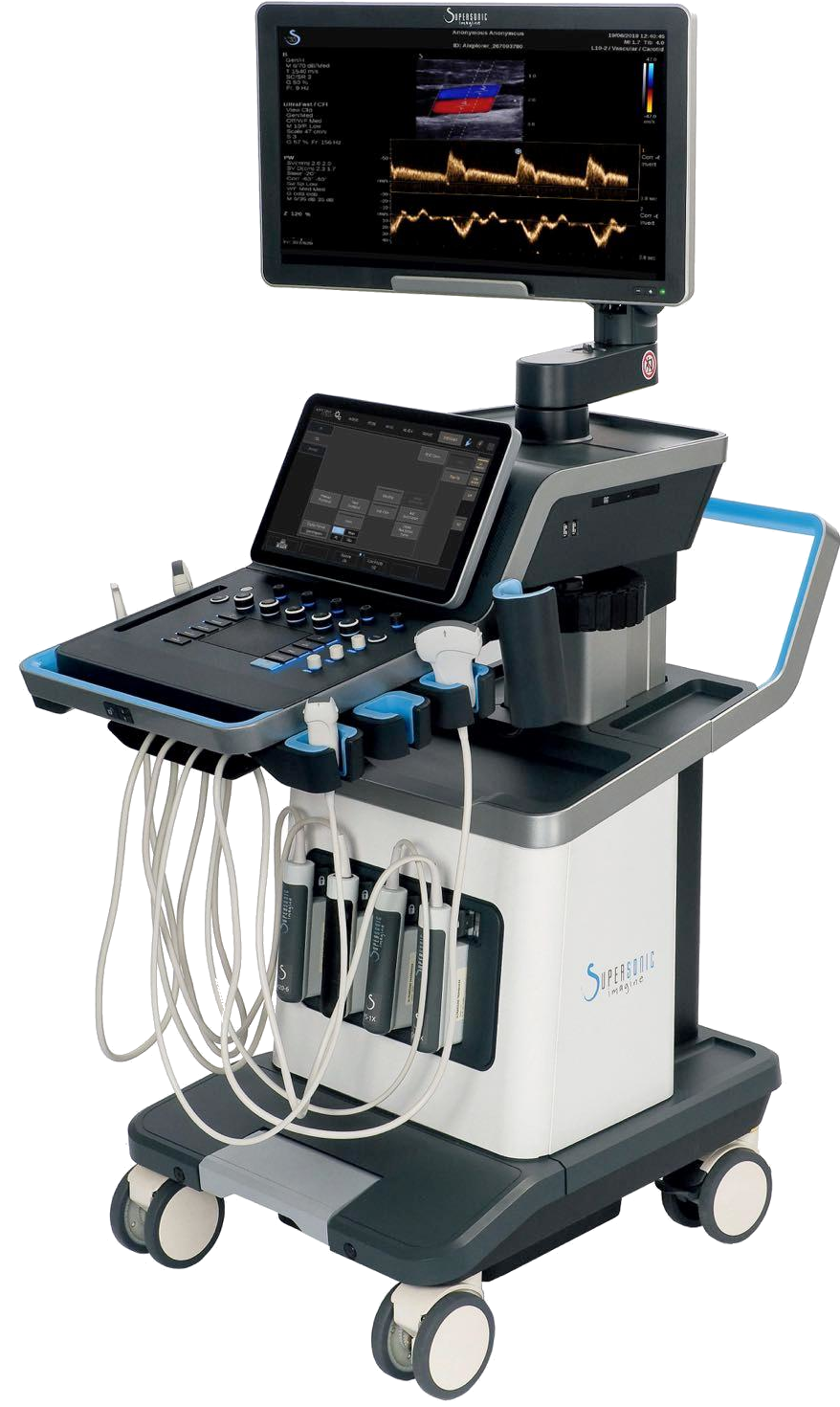
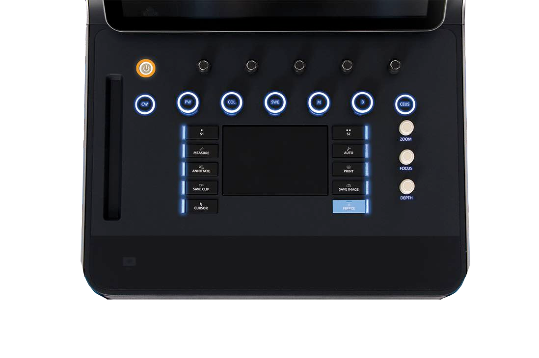
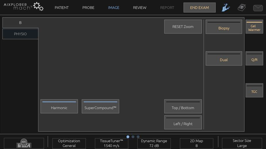
Ultrasound system interface
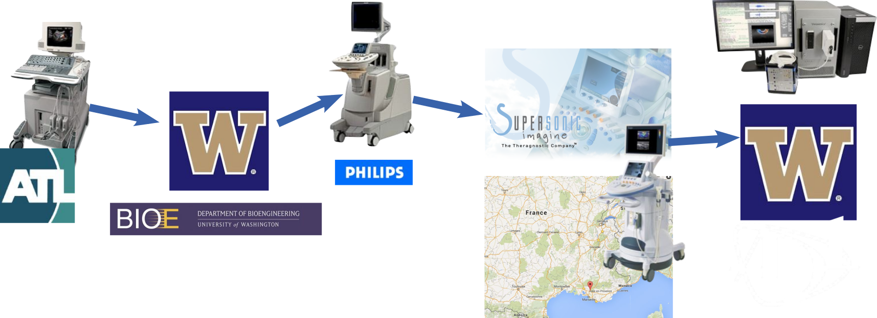
Ultrasound timeline
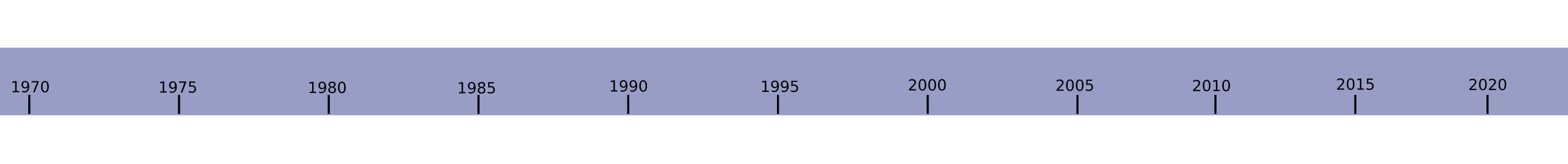 History of Ultrasound
Components of an ultrasound system
Components
- Transducer/connector
- Front end/Data acquisition board
- PCI express to computer
- More processing on GPU
- Display to monitor
- Power supply
Departments of an ultrasound company
- Hardware group
- Software group
- Ultrasound dev group
- Acoustic measurement group
- Clinical applications
- Marketing
- Sales
- Legal
- Administration
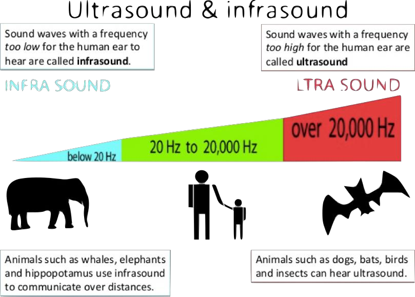
How does ultrasound work?
Echo location
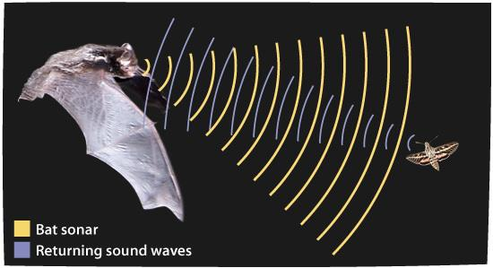
Ultrasound
Sound propagation in different medium
| Medium | Speed of sound | Wavelength (1 MHz) |
|---|---|---|
| Air | 300 m/s | 0.3 mm |
| Water | 1480 m/s | 1.48 mm |
| Tissue | 1540 m/s | 1.54 mm |
| Bone | 4000 m/s | 4.0 mm |
How do we generate ultrasound waves
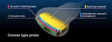
- Piezoelectric crystals
- Apply voltage
- Crystals vibrate at MHz frequencies
Principle of ultrasound image formation
Week 1: Wave propagation and ultrasound physics
- Wave equation.
- Description of wave propagation.
- Propagation in different materials.
- Time domain simulations.
Week 2: Diffraction and beams
- Integral solution of wave equation.
- Fourier analysis.
- Principles of diffraction.
- Relation of acoustic field and aperture function.
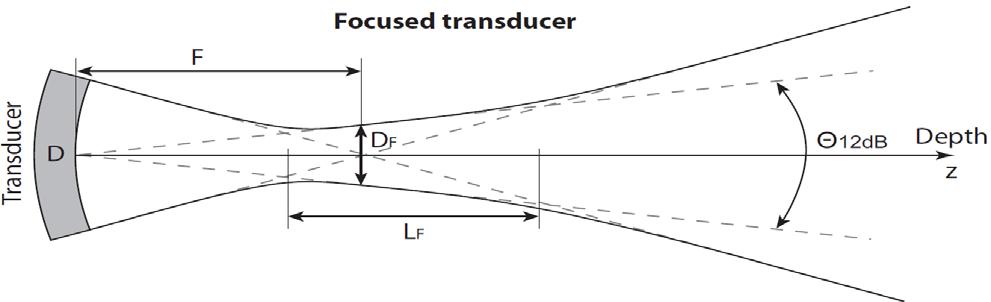
Week 3: Transducers/arrays and system architectures
Transducers and arrays
- Principles of electrical/acoustical transduction.
- Piezoelectric transducers.
- Transducer design and components.
- Transducer arrays.
System architectures
- "Conventional" system architecture and components.
- Modern system architecture.
- Portable/ultra-portable system.
Week 4: Signal processing tools
- Convolution.
- Fourier analysis.
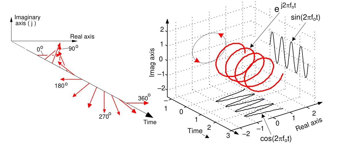
- Complex numbers.
- IQ and Hilbert transform.
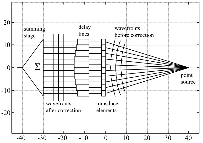
Week 5: Beam formation
- Diffraction equations for aperture.
- Transmit beamforming.
- Dynamic receive beamforming.
Week 7: B-mode imaging
- Ultrasound propagation in heterogenous media.
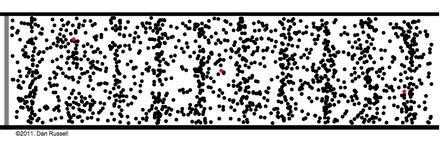
- Ultrasound scattering mechanisms.
- Principles of speckle.
- Real-time image reconstruction.
- Imaging controls and algorithms.
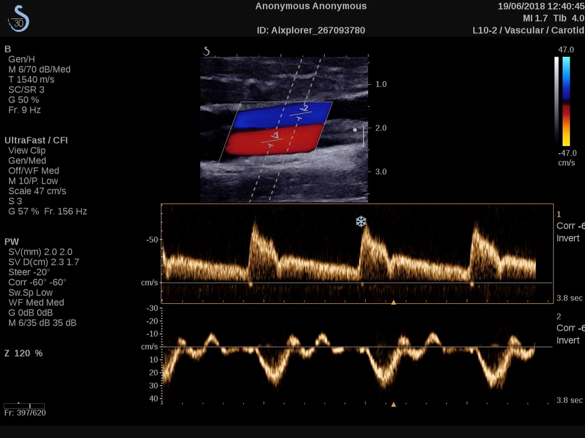
Week 8: Doppler Processing/imaging blood flow
- Basics of blood flow.
- Doppler physics.
- PW Doppler processing.
- Color Doppler processing.
- Plane wave Doppler imaging.
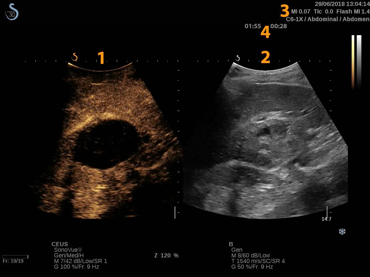
Week 9: Ultrasound contrast agents
- Introduction to microbubbles.
- Nonlinear response of microbubbles.
- Modes of oscillation.
- Imaging microbubbles.
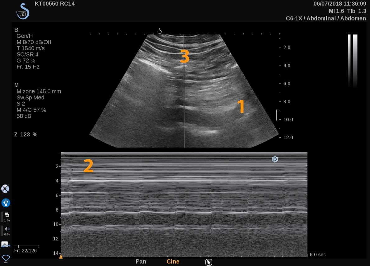
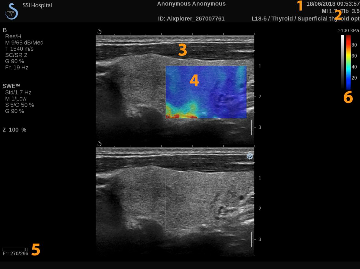
Week 10: Shear-wave Elastograpy
- Elastic properties of solids.
- Wave equation for elastic waves.
- Estimation of elastic modulus.
- Shear-wave imaging.
- Applications of SWE.
Week 11: Nonlinear/harmonic imaging
- Fundamentals of nonlinear propagation.
- Benefits of nonlinear imaging.
- Nonlinear scattering.
- Different approaches to imaging nonlinear echoes.