Vascular Research Page
![[PICTURE]](graft_banner.gif)
Arterial bypass grafts
Some of my current work is in the use of 3D ultrasound imaging for
surveillance of arterial bypass grafts in the lower limb. Another
topic of interest is measurment of abdominal aortic aneurysms. Below
are several examples of 3D reconstructions of vasculature.

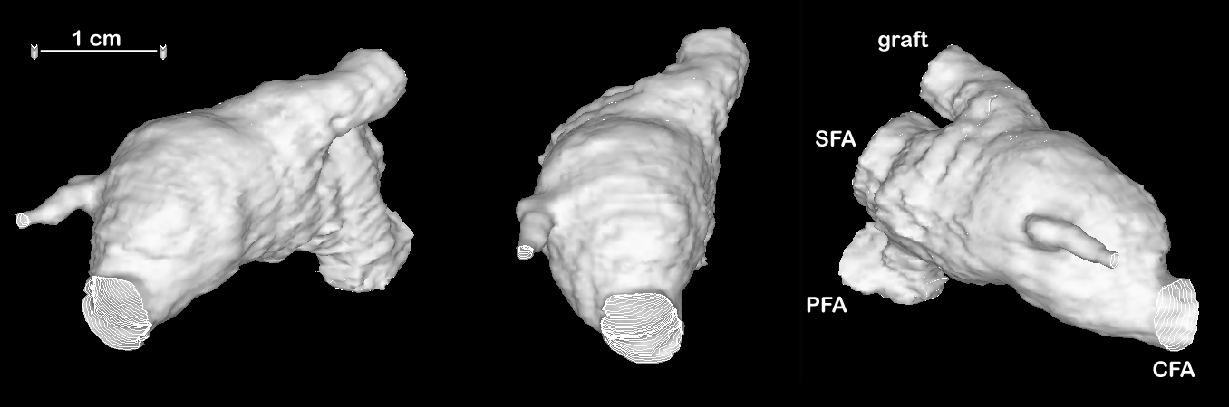
Three views of a surface rendering of a proximal anastomosis of a vein
graft, derived from direct volume reconstruction of 30 power Doppler
images. CFA: common femoral artery; SFA: superficial femoral artery;
PFA: profunda

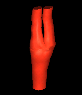
Surface reconstruction of the carotid bifurcation, generated from manual
outlines of B-mode images. normal volunteer
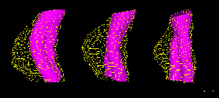
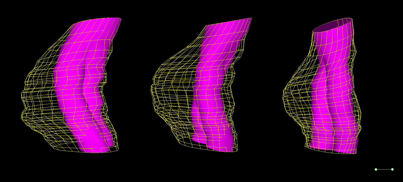
Abdominal aortic aneurysm (yellow mesh) repaired by an endovascular
stent graft
(purple surface). The surfaces were generated from manual outlines
of 30 B-mode images.
SPECTRAL WAVEFORM EXAMPLE
Leotta DF, Paun M, Beach KW et al.
"Measurement of Abdominal Aortic Aneurysms using
Three-Dimensional Ultrasound: Preliminary Report" J Vasc Surg,
33:700-707, 2001.
Abstract
Leotta DF, Primozich JF, Beach KW et al.
"Serial
Measurement of Cross-Sectional Area in Peripheral Vein Grafts using
Three-Dimensional Ultrasound" Ultrasound Med Biol, 27:61-68, 2001.
Abstract
Leotta DF, Primozich JF, Beach KW et al. "Cross-Sectional Area
Changes in Peripheral Vein Grafts Monitored by Three-Dimensional
Ultrasound Imaging" IEEE International Ultrasonics Symposium, San
Juan, Puerto Rico, October 2000.
Abstract
Leotta DF, Primozich JF, Beach KW et al. "Vein
Graft Surveillance using Three-Dimensional Ultrasound Imaging" SPIE Opto
Northwest, Bellevue, WA, November 1999.
Abstract
Daniel Leotta, PhD
Email: leotta@u.washington.edu
Web: http://staff.washington.edu/leotta/