Neuroscience For Kids
The Eye
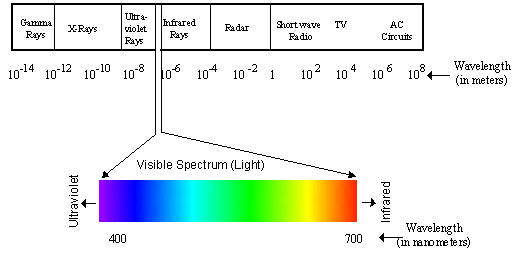
 Humans are very visual
animals...we use our sense of sight to interpret much of the world around
us. What we see is called "light." However, what we see is really only a
small part of the entire "electromagnetic spectrum." Humans can see only
the wavelengths of electromagnetic radiation between about 380 and 760
nanometers...this is light.
Humans are very visual
animals...we use our sense of sight to interpret much of the world around
us. What we see is called "light." However, what we see is really only a
small part of the entire "electromagnetic spectrum." Humans can see only
the wavelengths of electromagnetic radiation between about 380 and 760
nanometers...this is light.
 Our eyes do not have detectors for wavelengths of energy less than 380
or greater than 760 nanometers, so we cannot "see" other types of energy
such as gamma or radio waves. Rattlesnakes, however, can detect
electromagnetic radiation in the infrared range and use this ability to
find prey.
Our eyes do not have detectors for wavelengths of energy less than 380
or greater than 760 nanometers, so we cannot "see" other types of energy
such as gamma or radio waves. Rattlesnakes, however, can detect
electromagnetic radiation in the infrared range and use this ability to
find prey.
Electromagnetic Spectrum
Vision
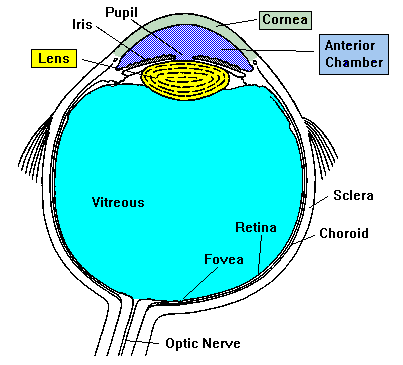 First, some specifics about the eye: the human eye is about 2.5 cm in
length and weighs about 7 grams. Light passes through the cornea, pupil
and lens before hitting the retina. The iris is a muscle that controls
the size of the pupil and therefore, the amount of light that enters the
eye. Also, the color of your eyes is determined by the iris.
First, some specifics about the eye: the human eye is about 2.5 cm in
length and weighs about 7 grams. Light passes through the cornea, pupil
and lens before hitting the retina. The iris is a muscle that controls
the size of the pupil and therefore, the amount of light that enters the
eye. Also, the color of your eyes is determined by the iris.
The vitreous or vitreous humor is a clear gel that provides constant pressure to maintain the shape of the eye. The retina is the area of the eye that contains the receptors (rods and cones) that respond to light. The receptors respond to light by generating electrical impulses that travel out of the eye through the optic nerve to the brain.
Six bands of muscles attach to the eyeball to control the ability of the eye to look up and down and side to side. These muscles are controlled by three cranial nerves. Four of the muscles are controlled by the oculomotor nerve (cranial nerve III), one muscle is controlled by the trochlear nerve (cranial nerve IV) and one muscle is controlled by the abducens nerve (cranial nerve VI.)
Parts of the Eye
- Aqueous Humor: Clear, watery fluid found in the anterior chamber of the eye.
- Choroid: Layer of blood vessels that nourish the eye; also, because of the high melanocytes content, the choroid acts as a light-absorbing layer.
- Cornea: Transparent tissue covering the front of the eye. Does not have any blood vessels; does have nerves.
- Iris: Circular band of muscles that controls the size of the pupil. The pigmentation of the iris gives "color" to the eye. Blue eyes have the least amount of pigment; brown eyes have the most.
- Lens: Transparent tissue that bends light passing through the eye. To focus light, the lens can change shape by bending.
- Pupil: Hole in the center of the eye where light passes through.
- Retina: Layer of tissue on the back portion of the eye that contains cells responsive to light (photoreceptors).
- Rods: Photoreceptors responsive in low light conditions.
- Cones: Photoreceptors responsive to color and in bright conditions.
- Sclera: Protect coating around the posterior five-sixths of the eyeball.
- Vitreous Humor: Clear, jelly-like fluid found in the back portion of the eye. Maintains shape of the eye.
 Hear IT! |
"Cornea" | "Iris" | "Retina" |
Learn More!
- Take a short on-line, interactive quiz about the eye.
- Complete a crossword puzzle about vision.
- Play an interactive word search game about the eye.
- Do You Wear Glasses?
- Experiments and Activities about Vision
- The retina
- Lesson Plan: The Eye
- Common Eye Disorders
- Eye Anatomy
Did you know?  |
15 million people in the United States have serious vision problems;
over 500,000 people in the US are blind. Eye injuries account for 4% of
the cases of blindness. Read more about eye
safety. |
Copyright © 1996-2021, Eric H. Chudler All Rights Reserved.